Product
Current location: Home Product New Energy SEM
  • Verios 5 XHR SEM ultra-high resolution scanning electron microscope
  • Verios 5 XHR SEM ultra-high resolution scanning electron microscope

Verios 5 XHR SEM ultra-high resolution scanning electron microscope

Characterize and analyze nanomaterials using sub nanometer resolution and high material contrast.
DetailsApplicationParameter

Verios 5 XHR SEM scanning electron microscope

The Verios 5 XHR SEM scanning electron microscope provides sub nanometer level resolution in the entire energy range of 1 keV-30 keV with excellent material contrast. Unprecedented automation and ease of use enable users of any level of experience to utilize this performance.

Experience the advantages provided by Verios XHR SEM scanning electron microscopy:

  • Using the industry's most advanced electronic source monochromator UC to achieve high-resolution nanomaterial imaging, sub nanometer performance can be achieved in the range of 1-30 kV.
  • Excellent performance of electron beam sensitive materials, with a landing energy of 20 eV and high sensitivity of detectors inside and under the lens, as well as signal filtering, can achieve ultra-high resolution and high signal-to-noise ratio images under low dose conditions.
  • With the Elstar electron beam barrel using SmartAlign and FLASH technology, it can greatly shorten the time for users with various levels of experience to obtain nanoscale information.
  • By utilizing ConstantPower lenses, electrostatic scanning, and the selection of two high-precision piezoelectric ceramic stages, consistent measurement results were obtained.
  • Equipped with a large chamber, the flexibility of accessories is high.
  • Use Thermo Scientific AutoScript 4 software (optional Python based application programming interface) to achieve unmanned and automated SEM operation of the instrument.


The main characteristics of Verios 5 XHR SEM scanning electron microscopy

SmartAlign technology

When using SmartAlign technology, users do not need to center the electron beam barrel in any way, which not only minimizes maintenance but also improves electron microscope productivity.

Innovative electronic optical systems

Including Thermo Scientific's patented UC (monochromator) electron gun, ConstantPower lens, and electrostatic scanning, accurate and stable imaging can be achieved.

Subnanometer resolution

Elstar Schottky electronic source monochromator (UC) FESEM technology and performance, achieving sub nanometer level resolution from 1 to 30 keV.

Consistent measurement results

Verios scanning electron microscopy is highly suitable for laboratory metrology applications and can calibrate NIST certified standards at high magnification.

Low dose operation and optimal contrast selection

By combining advanced high sensitivity, in tube and under lens detectors, and signal filtering functions, low-dose operation and optimal contrast selection can be achieved.

Easy access to beam landing energy

As low as 20 eV, extremely high resolution can also be achieved for true surface characterization.

Unmanned SEM operation

With the help of AutoScript 4 software, an optional Python based application programming interface (API) is available.

Large chamber

You can choose from two high-precision and stable piezoelectric ceramic driving sample stages.

Applications of Verios 5 XHR SEM Scanning Electron Microscopy
使用电镜进行基础材料研究

Basic material research

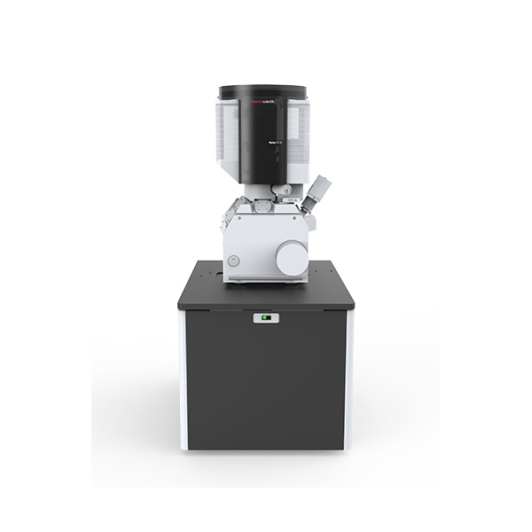
Research on new materials on an increasingly smaller scale to maximize control over their physical and chemical properties. Electron microscopy provides researchers with important insights into the properties of various materials at the micrometer to nanometer level.

半导体寻路

Semiconductor exploration and development

Advanced electron microscopy, focused ion beam, and related semiconductor analysis techniques can be used to identify feasible solutions and design methods for manufacturing high-performance semiconductor devices.

半导体良率提升和计量

Yield improvement and measurement

We provide advanced analytical capabilities for defect analysis, metrology, and process control, aiming to help increase productivity and improve the yield of a range of semiconductor applications and equipment.

半导体故障分析

Semiconductor fault analysis

The increasingly complex structure of semiconductor devices leads to more hidden defects caused by faults. Our next-generation semiconductor analysis workflow can help you locate and characterize subtle electronic issues that affect mass production, performance, and reliability.

物理和化学表征

Physical and chemical characterization

The sustained consumer demand has driven the creation of smaller, faster, and cheaper electronic devices. Their production relies on efficient instruments and workflows, enabling electron microscopy imaging, analysis, and characterization of various semiconductor and display devices.

specifications

Electron beam resolution

  • 0.6 nm at 30 kV STEM (optional)
  • 0.6 nm at 2-15 kV
  • 0.7 nm at 1 kV
  • 1.0 nm at 500 V

Standard detector

ETD, TLD, MD, ICD, beam current measurement, Nav Cam, IR cameras

Optional detector

Optional detectors | EDS, EBSD, RGB cathodoluminescence, Raman, WDS, etc

Carrier bias (beam deceleration, optional)

Standard configuration includes up to -4000 V

Sample cleaning

The standard configuration includes an integrated plasma cleaning device

Sample manipulation

Verios 5 UC

  • 5-axis electric common center loading platform, equipped with XYR axis piezoelectric drive.
  • XY range 150 x 150 mm270 ° tilt range.
  • Load through the door.

Verios 5 HP

  • Installed chamber, ultra stable 5-axis all piezoelectric driven loading platforms.
  • XY range 100 x 100 mm270 ° tilt range.
  • Load through automatic load lock.


chamber

379 mm inner diameter, 21 ports

Software Options

  • Thermo Scientific Maps software that uses partitioning and stitching for automatic large-scale collection; Related work
  • Thermo Scientific AutoScript 4 software; Application Programming Interface Based on Python
  • Pattern generation software
  • TopoMaps for image colorization, image analysis, and 3D surface reconstruction